Using photoluminescent nanorods as ultimate probes of fluid flow
19 June 2017
At HIMS Fred Brouwer, professor of Spectroscopy and Photonic Materials, together with research technician Michiel Hilbers contributed with confocal imaging and single-particle measurements of the nanorods. The collaboration was supported by LaserLab Europe.
Ultimate confirmation
The study of fluid flow in capillary networks is relevant to many fields. As an example, the determination of blood circulation in arteries is an important aspect of the study of plaque formation in atherosclerosis. Although hydrodynamic simulations can yield important information, experimental studies are needed for ultimate confirmation.
However, characterizing flows on the scale of a few hundred nanometers is rather difficult. The current technique of particle imaging velocimetry (PIV), tracking the displacements of fluorescent microspheres, cannot practically be used for local real-time observation of dynamic systems. Furthermore, in the case of velocity gradients (shear) common to capillary networks, PIV displays poor signal-to-noise ratios and scant spatial resolution.
Rods rather than spheres
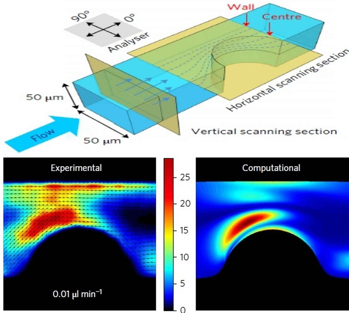
In their Nature Nanotechnology paper the French-Dutch research team now reports the use of nanorods rather than spheres. They show that instant detection of the collective orientation of the nanorods in a small focal volume enables the direct measurement and fast scanning of the local shear rate. As a proof of concept they demonstrate tomographic mapping of the shear distribution in a microfluidic model system using scanning confocal microscopy.
The researchers synthesized nanorods of lanthanum phosphate (LaPO4) crystals, doped with luminescent europium (Eu3+) ions. Like logs of trees floating on a river these nanorods, 10 nm in diameter and 200 nm in length, gradually orient themselves along the direction of flow. Thanks to the strongly polarized light emission properties of the europium ions their spatial orientation could be traced by means of their emission spectrum. Thus, it became possible to analyze, in real time and with unmatched resolution, the flow of a fluid in a small sized microfluidic channel.
This work opens promising perspectives for the fundamental understanding of phenomena related to the flow of a fluid in complex channels. Beyond this, these orientation probes could also be used in biology, to follow in-situ complex mechanisms related to the dynamics of orientation of bio-macromolecules in order to explain their properties and their modes of action.
Publication
JW Kim, S. Michelin, M. Hilbers, L. Martinelli, E. Chaudan, G. Amselem, E. Fradet, JP. Boilot, A.M. Brouwer, C. N. Baroud, J. Peretti, T. Gacoin: Monitoring the orientation of rare-earth doped nanorods for flow shear tomography Nature Nanotechnology, published online 19 June 2017. DOI: 10.1038/NNANO.2017.111